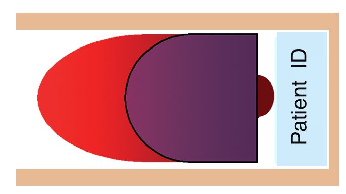
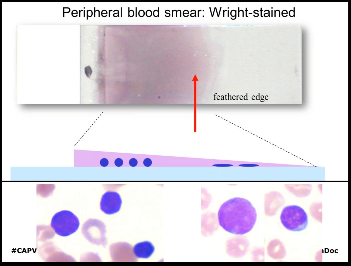
Feathered Edge Blood Smear Slide

Part One Deferential White Blood Cells Count Diff Wbcs Count And Microscopic Examination Of Well Stained Blood Film Page 2 Medicine Science And More
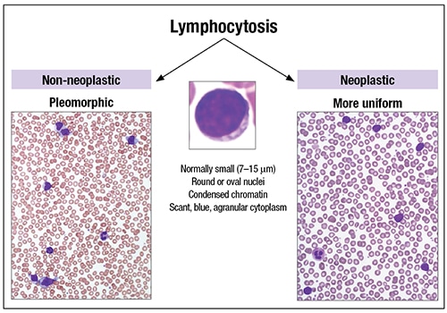
Evaluating A Blood Film Wbc Morphology Wbc Blood Parasites And Ppt Video Online Download

Diagnostic Blood Smear Preparation Clinician S Brief
Cvm Ncsu Edu Wp Content Uploads 16 09 Blood Smear Basics 16 Pdf

Smear Examination Eclinpath

Hematology Hemostasis College Of Veterinary Medicine At Msu
The size of the drop of blood and viscosity of the sample may lead to inconsistencies on the part of even the most practiced laboratorians.
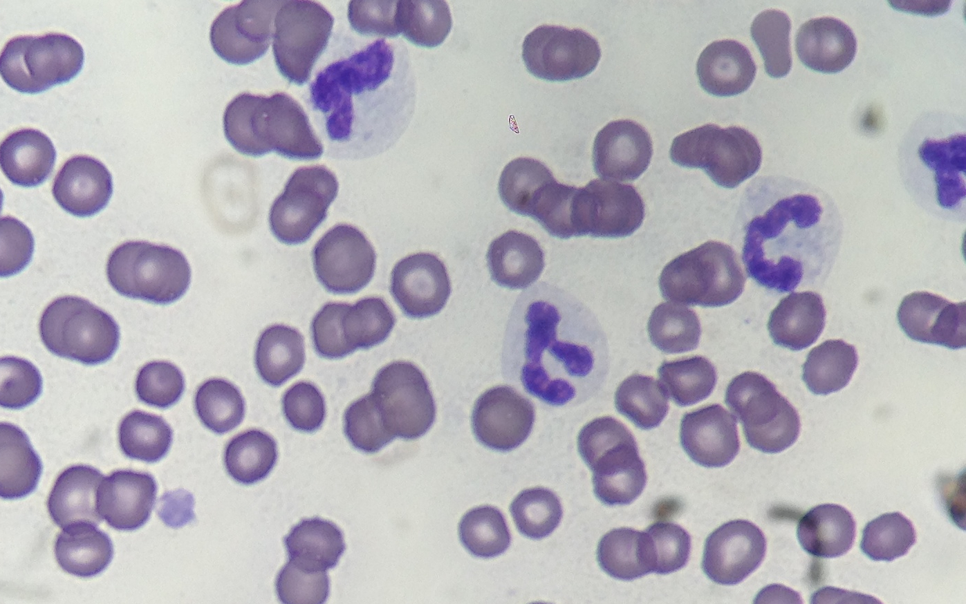
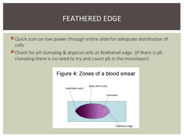
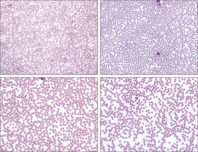
Feathered edge blood smear slide. Smear is spread across (side to side) both sides of the slide to the edge 5. Just give it a quick glance and make sure the red cells aren’t either piling up all over each other, or spread out too far with lots of holes in between – like the red cells in the image above. This is the part, where large particles are gathered, so it is useful to quickly check it, to detect some abnormalities like microfilaria, platelet clumps and unusual large cells.
Don’t hesitate with the spreader slide. The slide in your hand is the spreader slide. Rainbow sheen at end of the slide (feathered edge and monolayer) 6.
Knowing where to look on the slide can make all the difference between being able to see what's there, and frustration. The smear should cover 1⁄ 2-3⁄ 4 of the slide and finish with a “feathered” edge. Push the second slide back until it contacts the drop of blood.
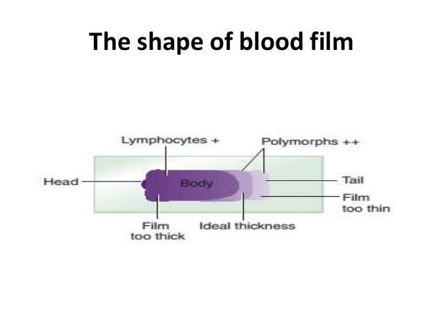
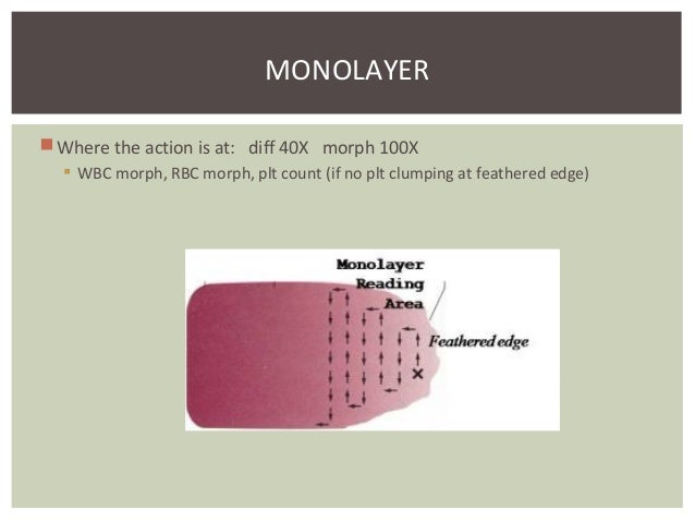
Zones of a blood smear. Don’t go too fast. The pale middle band of the gradient is the monolayer.
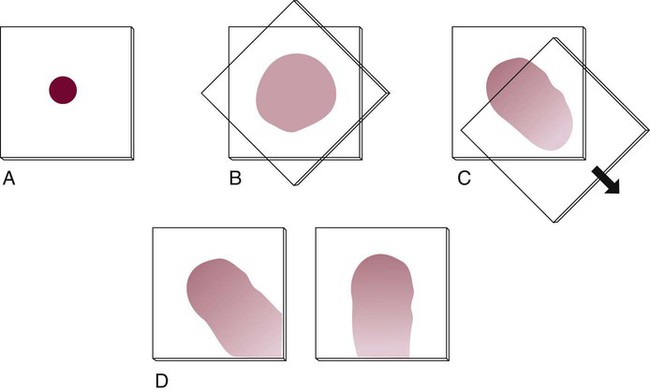
Common causes of a poor blood smear 1. The monolayer will contain cells that are the easiest to identify and are the least distorted. Using your dominant hand, place the edge of the other slide at an approximately 35-45⁰ angle on the first glass slide, in front of the blood drop.
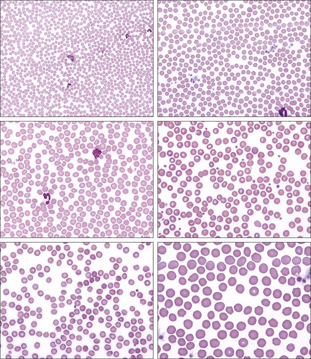
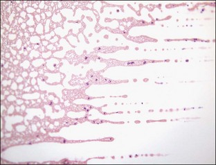
These pictures show the low and high power appearance of the feather edge, which is too thin to use for identification of leukocytes and assessment of morphologic too thin abnormalities. Take a second slide and lie the edge flat on the smear slide. You want to be way out on what we call the 'feathered edge' of the slide.
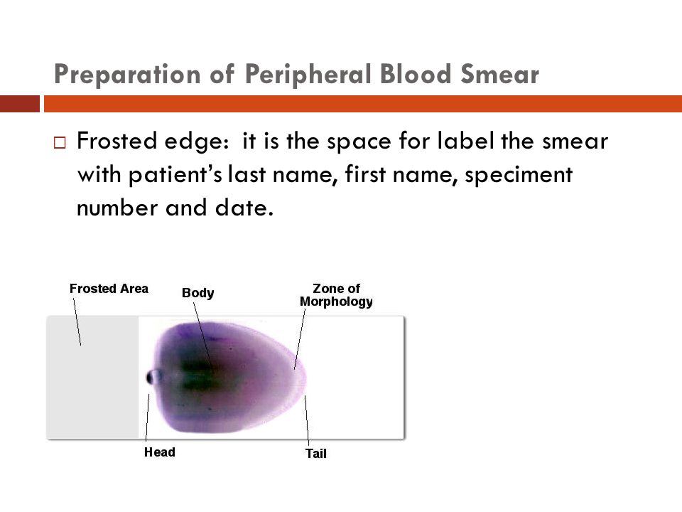
Slide is labelled with the patient identifier and type of sample (in this case, “blood film”). Once the smears are dry, place them in the cardboard slide holders, tape shut, and transport to ALI along with the EDTA blood. Drop of blood too large or too small.
Troubleshooting blood smear errors Problem Solutions Short smear • Use a larger droplet of blood. The smear should have an even "feathered edge." Allow the smears to air dry completely. This technique requires at least two 3 × 1-inch (75 × 25-mm) clean glass slides.
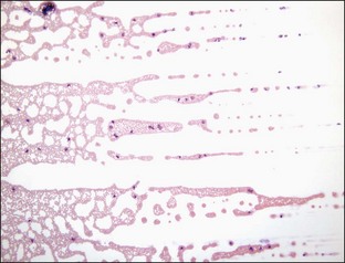
-leukocyte distribution at feathered edge allows for the identification of abnormal cells that tend to locate on the edges of the smear disadvantages of wedge smear poor leukocyte distribution with monocytes and neutrophils being drawn out of the optimal counting area to the feathered edge acceptable samples for peripheral blood smear. Once the blood smear is stained, the cells are visually inspected with a microscope. View of the monolayer and feathered edge at 100X oil immersion resolution.
Submit 1 well-prepared, thin blood smear on clean, grease-free slide. This is the very end area of the smear and consists of a monolayer of cells. Whereas a smaller angle gives a thin smear.
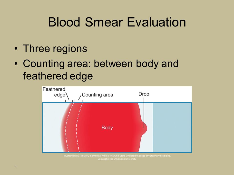
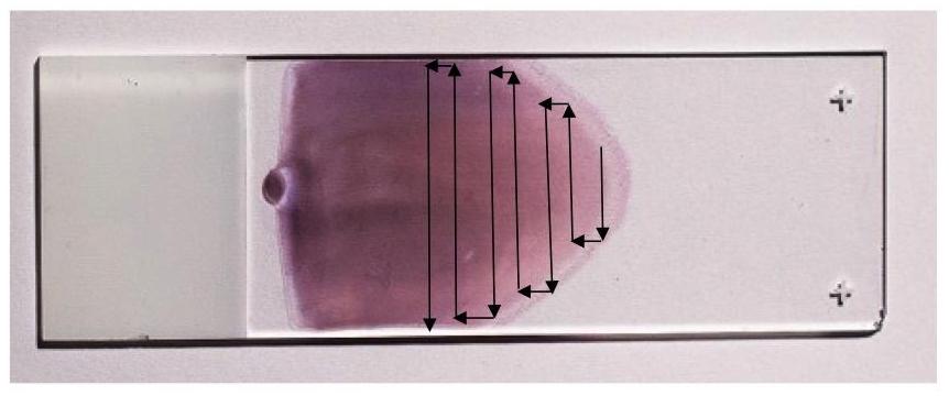
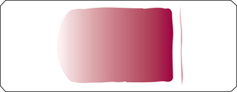
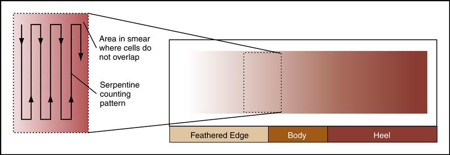
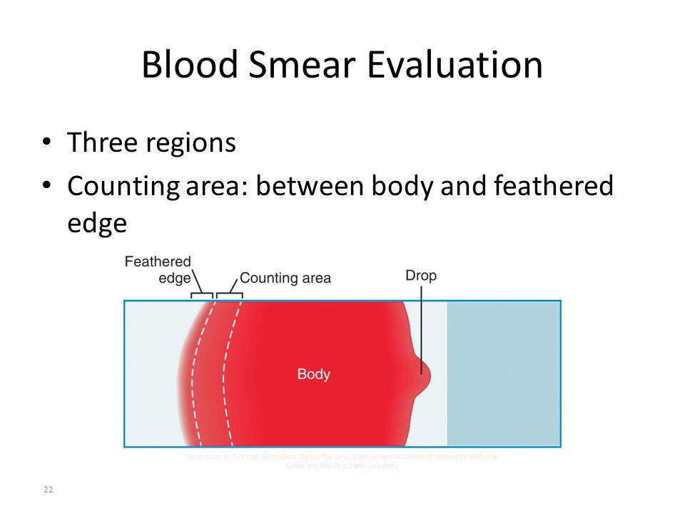
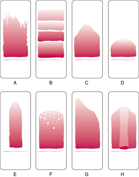
Blood smears have three major areas:. This method produces a gradual decrease in thickness of the blood from thick to thin ends with the smear terminating in a feathered edge approximately 2 mm long. And (3) the feathered edge (the most external area).
Label slide with patient’s full name (first and last) and date and time of. Under 100x oil immersion magnification, platelets on each of 10 fields are counted, averaged, and multiplied by 15.000 in order to get the estimated number per /µl. Proper blood smear preparation.
Blood Smear 6a 6b 7 The PULL technique:. Smears may be made using coverslip or push-smear slide techniques. Failure to keep the spreader slide at a 30° angle with the slide 13.
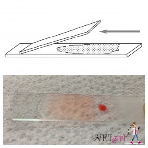
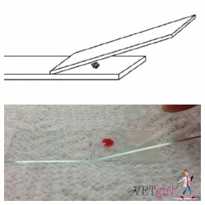
With its fully digital, automated scan and image acquisition system, the Full Field PBS offers unique user experience of in-slide navigation to specified locations within a slide, all through a. Using a capillary tube, place a 2-3mm drop of blood about 1cm from the frosted end of a clean slide that is on a flat surface. If the smear is prepared from an EDTA sample, the specimen should be free of clots.
• Increase the speed of the spreader slide. Don’t use a dirty or chipped spreader slide. The wedge smear is a convenient and commonly used technique for making peripheral blood smears.
Feathered edge of a blood smear. Allow the blood to spread along the edge of the slide. Allow slides to dry on a flat surface.
Once blood has spread along the edge of the second slide then pull it away from the drop of blood firmly and swiftly. Fresh smears are particularly important when evaluating blood smears for:. Long smear / no feathered edge • Use a smaller drop of blood.
This slide is held at an angle (about 30 degrees) and drawn backward into the drop of blood. If using a needle and syringe, first remove the needle and then touch the end of the syringe to the slide. ADVANCE Staff November 7, 13 February 13, 19.
Make sure that the smears have a good feathered edge. Quickly push the upper (spreader) slide toward the unfrosted end of the lower slide. You should be a couple medium-power fields in from the “feather edge,” which is the thin edge of the smear where the cells are all spread out and there are huge empty spaces.
The smear is rapidly air dried, labeled in pencil on the frosted end, and stained. If there is a thick line of blood where the slide stopped, it’s an indication of a poorly made smear. Don’t use a drop of blood that is too small.
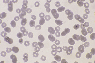
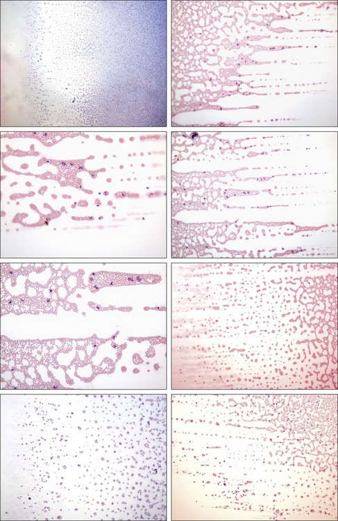
The smear is greater than 25 mm long and the feathered edge stops approximately 10 mm from the end of the slide. The loss of central pallor is likely secondary to thinning of the blood smear at the feathered edge, and leukocyte distortion is likely secondary to dragging of cells to the feathered edge that can occur during slide preparation. Morphologic evaluation of cells Organism identification High quality slides should have:.
Place a drop of blood approximately 4 mm in diameter on the slide, approximately 0.5 cm from the frosted area. Furthermore, the technology utilizes monolayer identification in support of long and short smears and automates the analysis process by pre-classifying 0 white blood cells. This allows clinical laboratories the ability to capture digital scans with full field view of the the monolayer and feathered edge at 100X oil immersion resolution level.
The smear itself should look very smooth with a seamless progression to what is called a “feathered edge”. • Decrease the angle of the spreader slide. It features adaptive monolayer identification for optimal imaging and analysis of short and long smears, including the feathered edge of the sample.
Push the spreader slide smoothly and quickly down the slide producing a feathered edge. With a lead pencil, label the slides with the. Macroscopically, the smear should exhibit a gradual transition from thick to thin, ending with an acceptable feather edge free of streaks, waves, or holes.
Using gentle pressure, gently pull the second slide back into the blood drop and allow the blood to spread to the edge of the slide. Allow the smear to air dry. (if there are platelets clumping there is no need to try and count platelets in the monolayer).
Tail edge, or feathered edge of smear. A well-developed feathered edge. - pusher slide backs into drop of blood and pushes it forward - angle of pusher slide:.
More HORIZONTAL -- long, thin smear and no feathered edge - use clean edge of pusher - want 45 degree angle and push forward at EVEN speed. A thin monolayer region where erythrocytes and leukocytes can be adequately evaluated. Closeups of the feathered edge of blood smears.
Unstained and stained blood smears with a near perfect “feathered edge” are shown in Figure 4. As the blood spreads along the trailing edge of the spreader slide, this slide is advanced in a rapid and smooth motion, carrying the blood with it. More VERTICAL -- short thick smear - angle:.
High-quality, beveled-edge microscope slides are recommended. Labs don’t report bands. This may be done by hand or using an automated slide maker coupled to a hematology analyzer.
For best results use a Bev-L-Edged slides for both the blood smear & second slide (“spreader”). The spreader slide is advanced forwards, creating a smear with a feathered edge If too much blood is applied to the slide or taken up by the spreader slide the smear will be too long and the cells will be pushed over then end of the slide. It's easy to run it through a slide.
Not a good area to review red cell morphology. Blood smear preparation is a technical skill that requires practice. A feathered edge and extend 1/2 to 2/3 the length of the slide.
Scopio Labs Receives FDA Clearance for its AI-Powered Full Field Peripheral Blood Smear (Full Field PBS) Application. (1) the thick inner area (body);. This region should be noticeably thinner than the body, but should blend in with the body of the smear.
A blood smear is prepared by placing a drop of blood on a microscope slide and using a second slide held at an angle to spread the blood and pull it across the slide, forming a "feathered edge" consisting of a single layer of cells at the end of the smear. Push-smear techniques result in a more consistent "feather-edge" and a better monolayer (red cell area) for. View chapter Purchase book.
That is, you want to be in the shallow end of the blood smear, not in the middle of the slide. But it also raises questions of accuracy. A monolayer area just behind the feathered edge.
Feathered edge of peripheral blood smear 6 years ago by Medical Labs 0 Quick scan on low power through entire slide for adequate distribution of cells then Check for platelets clumping & atypical cells at feathered edge. Feathered edge of peripheral blood smear Tweet Quick scan on low power through entire slide for adequate distribution of cells then Check for platelets clumping & atypical cells at feathered edge. Draw the spreader slide rapidly and smoothly over the entire length of the smear slide pulling a thin even film behind it.
This should be the first part of the smear that is examined at low power to detect platelet clumps and microfilaria, but should be avoided when evaluating blood cells at higher power. Blood smears Troubleshooting blood smears Do make sure there is a feathered edge. Blood smears are notable for numerous target cells that are relatively uniform in appearance.
One slide serves as the blood smear slide and the other as the spreader slide. • Decrease the speed of the spreader slide. A platelet count can be estimated from a monolayer blood smear, if there are not platelet clumps along the feathered edge.
Prepare a second slide. This part of the smear should be scanned at low power to detect platelet clumps and microfilaria, as shown in the top panel, but should be avoided when using the oil immersion objective. Other variables (hematocrit of the sample, wedge slide feathered edge, staining technique, humidity, human factors, etc.) suggest that these manual counts.
Coverslip smears result in more equitable distribution of leukocytes. • Increase the angle of the spreader slide. Place a small drop of blood on the pre-cleaned, labeled slide, near its frosted end.
The feathered edge This is the end of the blood smear and should be completely present and fully stained (a smear that is “too long” will lack a feathered edge). Spreader slide pushed across the slide in a jerky manner. This edge should have a fine, feathery appearance;.
The fixative is essential for good staining and presentation of cellular detail. The inner area is the thickest area of the smear, and cells are usually too contracted, distorted, or poorly stained for reliable evaluation. Prepare with a “feathered edge.” Smear should be no more than a single cell thick.
Pick up a second clean slide and hold it by placing your first two or three fingers on one edge of the slide and your thumb on the opposite edge;. To be able to see individual cells, it is first necessary to create a very thin film of blood with a “feathered edge” (a single layer of cells) on a glass slide. The desired and only lab-accepted "smear" results in a feathering of the blood, or a increasingly thinning of the amount of blood across the plate, in turn creating a feathered appearance of the.
The resulting blood smear should be at least two-thirds of the length of the slide. Microscopically, there should be a uniform distribution of platelets without clumping. The slide is left to air dry, after which the blood is fixed to the slide by immersing it briefly in methanol.
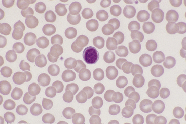
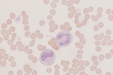
However, some large cells such as blasts may roll to the edge of the smear during preparation and may be more readily found there on visual scanning of the smear. The best area for cell counting is the monolayer of cells (counting area) between the thick region and the feathered edge (Thrall et al., 04). Larger cells, such as this immunoblast (black open arrow), are often pulled to the feather edge in the preparation of blood smears as.
The end of smear is called a feathered edge and can be recognized by loss of erythrocyte central pallor and a reticular pattern of erythrocyte distribution. While the goal is to have the smear cover approximately two-thirds of the slide with a feathered edge at the end, the slightest adjustment of the hands vs. It is important to scan the feather edge of a smear during manual morphologic review to detect cells and even microfilarial worm s that have been "dragged" to this portion of the blood smear.
The blood film occupies the central portion of the slide and has definite margins on all sides that are accessible to examination by oil immersion. Left to air-dry and fixed with a quick dip into 100% alcohol, the appearance of a ‘feathered edge’, where blood cells are evenly distributed and only one cell thick, will prove the success of your blood smear technique. And quickly pushed forward to the end of the first slide.
Bring another slide at a 30-45° angle up to the drop, allowing the drop to spread along the contact line of the 2 slides. The blood will spread behind the spreader slide by capillary action and should be allowed to spread the full width of the spreader slide.
How Do I Make A Good Blood Smear Vetgirl Veterinary Ce Podcasts

Pin On Vet Stuff

Erythropoiesis Normal And Abnormal Barrett W Dick M D Ppt Video Online Download

How To Get The Most From Blood Samples A Guide To Producing Diagnostic Blood Smears The Veterinary Nurse
Cvm Ncsu Edu Wp Content Uploads 16 09 Blood Smear Basics 16 Pdf
Http Williams Medicine Wisc Edu Blood Smear Pdf

Blood Smear No Feathered Edge From Vetstream Definitive Veterinary Intelligence
Examination Of The Peripheral Blood Film And Correlation With The Complete Blood Count Oncohema Key
Three Minute Peripheral Blood Film Evaluation Preparing The Film Dvm 360
Http Williams Medicine Wisc Edu Blood Smear Pdf
Practical Challenges In Peripheral Blood Smear Evaluation Cap Today

Blood Smear Features Eclinpath

In Clinic Hematology The Blood Film Review Today S Veterinary Practice
How Do I Make A Good Blood Smear Vetgirl Veterinary Ce Podcasts

Feathered Edge Of Peripheral Blood Smear Medical Laboratories

How To Get The Most From Blood Samples A Guide To Producing Diagnostic Blood Smears The Veterinary Nurse
Blood Smear Technique For Veterinarians Agriculture And Food

Did You Know Platelet Clumping
Blood Smear Evaluation
Q Tbn 3aand9gctspsjesswmjwhmg3e8sztosard6m6lzpltqindfj5zodaggvyn Usqp Cau

Human Blood Film Slide Smear Wright S Stain Carolina Com
Peripheral Blood Smears Veterian Key
Cvm Ncsu Edu Wp Content Uploads 16 09 Blood Smear Basics 16 Pdf

Blood Film Wikipedia
Blood Smear Evaluation

Peripheral Blood Smears Veterian Key

Did You Know Platelet Clumping

In Clinic Hematology The Blood Film Review Today S Veterinary Practice
How Do I Make A Good Blood Smear Vetgirl Veterinary Ce Podcasts
Q Tbn 3aand9gcrupkzmxcqbtkvo1zut3ts7 C Cigmjwjgc Mfvwij5pzkzbp 8 Usqp Cau
Q Tbn 3aand9gcs Zmr Leol5dxxge7pdpmbwcvjnj G Tf10qzpbccvui5thu Usqp Cau

Blood Smear Platelet Evaluation Interpretation Clinician S Brief
1 General Assessment Veterian Key

Did You Know Platelet Clumping

Feathered Edge Of Peripheral Blood Smear Medical Laboratories
Cvm Ncsu Edu Wp Content Uploads 16 09 Blood Smear Basics 16 Pdf
Http Williams Medicine Wisc Edu Blood Smear Pdf
Blood Smear Preparation And Staining
Cvm Ncsu Edu Wp Content Uploads 16 09 Blood Smear Basics 16 Pdf

The Criteria Of A Good Blood Film Medical Laboratories
Cvm Ncsu Edu Wp Content Uploads 16 09 Blood Smear Basics 16 Pdf

Making A Great Blood Smear Sonopath

Blood Smear Platelet Evaluation Interpretation Clinician S Brief
1 General Assessment Veterian Key

Did You Know Platelet Clumping
Q Tbn 3aand9gcqfe1ahl4izyyvcdfxvjrvtdqt Ottsgewsz8 Ydwlabockagvh Usqp Cau

Tips For Making A Good Blood Smear Medical Laboratories
Blood The Good The Bad And The Ugly Microbiology A Laboratory Experience
Cvm Ncsu Edu Wp Content Uploads 16 09 Blood Smear Basics 16 Pdf
Camlt Org Wp Content Uploads 17 09 Hematology Essentials A Foundation For Accurate Smear Reviews 3 17 Pdf
Cvm Ncsu Edu Wp Content Uploads 16 09 Blood Smear Basics 16 Pdf

White Blood Cell Differential Wikipedia

How To Read A Blood Smear Pathology Student
Www Agric Wa Gov Au Sites Gateway Files Master your blood smear technique Pdf

Few Qrbcs Seen Black Circle In The Feathered Edge Area Of The Blood Download Scientific Diagram
Cvm Ncsu Edu Wp Content Uploads 16 09 Blood Smear Basics 16 Pdf

Blood Smear Authorstream
Cvm Ncsu Edu Wp Content Uploads 16 09 Blood Smear Basics 16 Pdf
Introduction To Peripheral Blood Smear Examination Oncohema Key

Feathered Edge Of A Blood Smear Eclinpath
Peripheral Blood Smears Veterian Key

What Are The Differences And Similarities Between Blood Film Test And Full Blood Count Quora
Cvm Ncsu Edu Wp Content Uploads 16 09 Blood Smear Basics 16 Pdf
Peripheral Blood Smears Veterian Key
Michael Arnold Md Phd Where You Look On The Slide Of A Peripheral Smear Can Change The Morphology Great Capvirtualpath Tip From Kmirza
Cvm Ncsu Edu Wp Content Uploads 16 09 Blood Smear Basics 16 Pdf

Diagnostic Blood Smear Preparation Clinician S Brief
5 Hematology Nurse Key

Blood Smear And Cell Breakthrough The Gulls Of Appledore

Practical Assessment Of Blood Smears In Dogs And Cats In Practice

In Clinic Hematology The Blood Film Review Today S Veterinary Practice

Blood Film Wikipedia
Week Four Hematology Cbc Leukocytes Ppt Video Online Download

How To Get The Most From Blood Samples A Guide To Producing Diagnostic Blood Smears The Veterinary Nurse
Cvm Ncsu Edu Wp Content Uploads 16 09 Blood Smear Basics 16 Pdf

In Clinic Hematology The Blood Film Review Today S Veterinary Practice
Blood Smear A Nada Al Juaid Ppt Video Online Download

In Clinic Hematology The Blood Film Review Today S Veterinary Practice
1 General Assessment Veterian Key

Hematology Hemostasis College Of Veterinary Medicine At Msu

In Clinic Hematology The Blood Film Review Today S Veterinary Practice
Peripheral Blood Smear Examination

Blood Smear Preparation Clinician S Brief

Practical Assessment Of Blood Smears In Dogs And Cats In Practice

Figure 1 From Automatic Zone Identification In Blood Smear Images Using Optimal Set Of Features Semantic Scholar
Cvm Ncsu Edu Wp Content Uploads 16 09 Blood Smear Basics 16 Pdf
1 General Assessment Veterian Key

Making A Great Blood Smear Sonopath
Ieeexplore Ieee Org Iel7 Pdf
Http Williams Medicine Wisc Edu Blood Smear Pdf

Examination Of The Peripheral Blood Film And Correlation With The Complete Blood Count Rodak S Hematology Clinical Principles And Applications 5th Ed
Introduction To Peripheral Blood Smear Examination Oncohema Key
1 General Assessment Veterian Key
Practical Challenges In Peripheral Blood Smear Evaluation Cap Today
Blood Smear Evaluation
Cvm Ncsu Edu Wp Content Uploads 16 09 Blood Smear Basics 16 Pdf
Cvm Ncsu Edu Wp Content Uploads 16 09 Blood Smear Basics 16 Pdf
Http Williams Medicine Wisc Edu Blood Smear Pdf


